Reproduction and Development
|
|
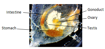
E. diaphanis showing reproductive organs.
Information from Ruppert et al. (2004).
|
E. diaphanis, like most ascidians, are simultaneous hermaphrodites. Self-fertilisation is, however, rare for the majority of ascidian species (Ruppert et al., 2004). The gonads, a testis and ovary, are located in the gut loop. The separate oviduct and sperm duct run parallel to the intestine and enter the atrium close to the anus (Ruppert et al., 2004). E. diaphanis is viviparous with embryological development occurring within the zooid. The eggs are gestated in the oviduct and the tadpole larvae are released.
|
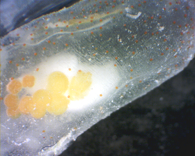
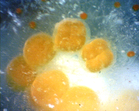
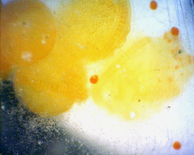
Images displaying embryo development in E. diaphanis. From let to right: developing embryos in the oviduct,
early emybrogenesis showing progressive development and cleavage, later development of the embyro dorsal
chord becoming visible.
|
Cleavage is holoblastic and bilateral with highly determinate development (Ruppert et al., 2004). The posterior end is marked by the blastopore which closes as the embryo elongates. The archenteron gives rise to the notochord and propergates the development of a band of mesodermal cells along the sides of the body. There is no mesodermal segmentation within ascidian larvae (Ruppert et al., 2004). The ectoderm differentiates as the nueral plate, sinks inward, and rolls upward forming the neural tube. Development eventually leads to a lecithotrophic tadpole larva. The larval life stage is usually short, less than 36hrs (Ruppert et al., 2004).
Tadpole larva and metamorphosis
The ascidian tadpole larva express all key chordate traits, but unlike the ancestral chordate it does not feed (Ruppert et al., 2004). In the absence of feeding the larval gut and associated structures may be non-functional and undifferentiated, or differentiated forming structures that will only function after metamorphosis (Ruppert et al., 2004).
The tadpole body is divided up into a visceral trunk and a locomotory tail. The trunk contains the cerebral vesicle and viscera. The tail is elongated with swimming musculature with continuous dorsal and ventral fins, the notochord and the dorsal neural tube (Ruppert et al., 2004). The tail is lost during metamorphosis. Structures such as the pharynx, siphons and digestive system are present but non-functional due to the larval cuticle covering the siphon openings (Ruppert et al., 2004). In E. diaphanis the embryo and larvae contain orange pigment (Fox, 2007).
Upon settlement, the larvae attaches to the substrate with three adhesive papillae (Ruppert et al., 2004). The larvae undergo a rapid metamorphosis into a juvenile zooid (Pechenik, 2010). During this metamorphosis the tail is retracted and absorbed, there is rapid growth of the inhalant siphon, the body rotates 90°, the atrium expands, the number of gill slits increases and the larval cuticle is molted (Ruppert et al., 2004).
Budding
Colonial growth occurs by budding. In general, zooids developing from a metamorphosed tadpole is called an oozooid and those formed by budding are known as blastozooids (Ruppert et al., 2004). The oozoids generally do not become sexually mature, therefore, the blastozooids are responsible for sexual reproduction (Ruppert et al., 2004).
|